Positron emission tomography (PET scan) is one of the most advanced imaging tests available at California Cardiac Institute. It maps your heart in remarkable detail, identifying blockages, reduced blood flow, and damage to the heart muscle. This noninvasive test helps us detect problems early and guide the right treatment plan.
Detect Blood Flow and Heart Muscle Damage
Positron emission tomography (PET scan) is a powerful, noninvasive imaging test that shows how well your heart works. By using a small amount of radioactive tracer, the scan highlights areas of your heart with reduced blood flow or damaged muscle. These allow our doctors to see blockages, evaluate heart function, and detect early signs of coronary artery disease, heart failure, or cardiomyopathy. Because it provides both structural and functional information, a cardiac PET scan in Los Angeles offers greater accuracy in diagnosing and monitoring heart conditions.
Positron Emission Tomography (PET Scan) Identifies:
- Blockages or narrowing in the coronary arteries
- Reduced blood flow to areas of the heart
- Damage to the heart muscle after a heart attack
- Changes in the structure of the heart muscle (cardiomyopathy)
- Early signs of heart failure or impaired function
- Effectiveness of treatments such as bypass surgery or angioplasty
Why is Positron Emission Tomography Done
A positron emission tomography (PET scan) is performed to give physicians a clear picture of how your heart is functioning at a cellular level. It helps detect reduced blood flow, heart muscle damage, and early signs of disease that may not appear on other imaging tests. This scan is often recommended when symptoms or risk factors suggest underlying heart problems.
You may need a PET scan if:
- You experience chest pain or shortness of breath
- You have risk factors for coronary artery disease
- You need an evaluation after a heart attack
- You are being monitored for heart failure or cardiomyopathy
- You require an assessment of how well treatments are working
What to Expect During Your PET Scan
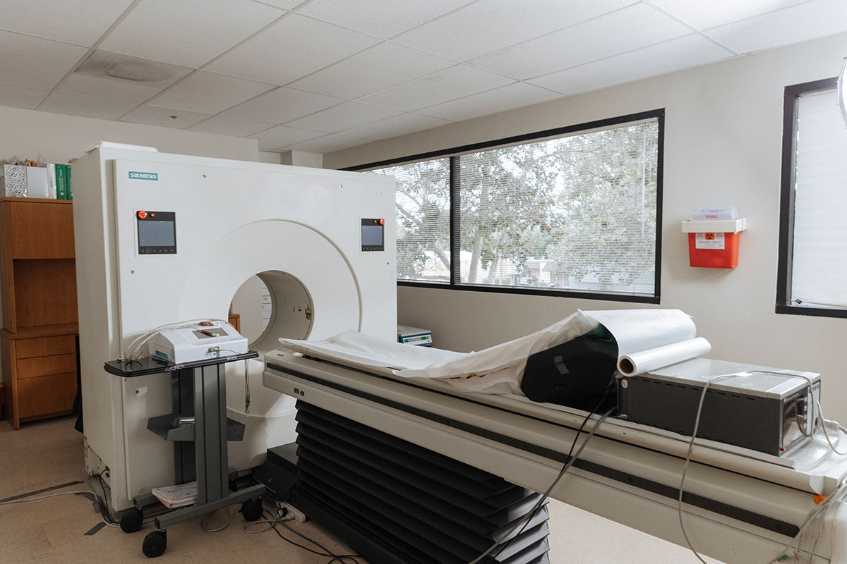
Before your PET scan, you may be asked to avoid eating for several hours to ensure accurate results. Once you’re ready, we insert a small IV line in your arm to deliver a radioactive tracer, usually a glucose-based compound. Because heart muscle cells use glucose for energy, this tracer shows us how much energy different areas of your heart are consuming. Areas with reduced blood flow or damaged tissue absorb less tracer, which becomes visible on the scan. You will lie flat on a cushioned table that slides into the PET scanner. The scanner detects tiny particles released by the tracer and uses this data to create highly detailed, 3D images of your heart. The test takes about 30 minutes, during which we ask you to remain still for the clearest results. Afterward, the tracer leaves your body naturally through urine.
Interpreting Your PET Scan Results
After your PET scan, our physicians carefully analyze the images to evaluate how well your heart is functioning. We look for areas where the tracer uptake is lower than normal, which may indicate reduced blood flow, scar tissue from a previous heart attack, or weakened heart muscle. These help us determine whether your heart muscle is still alive and viable. Once we interpret your results, we use this information to guide your care. If we see blockages, we may recommend further testing or procedures such as angiography. If the scan shows weak heart muscle, we tailor treatment to improve its function and prevent worsening heart failure. We use your results to design a personalized care plan focused on preventing complications, improving your quality of life, and promoting long-term heart health.

Schedule Your Consultation
California Cardiac Institute is committed to helping you live longer, healthier, and stronger by combining advanced medicine with preventive care. Our state-of-the-art imaging center includes a cardiac PET scan in Los Angeles, which maps your entire heart for accurate diagnosis. We offer same-day appointments and accept walk-ins, because heart conditions cannot wait. Our physicians use this powerful tool to detect disease early, guide treatment, and support your long-term health. Schedule your PET scan today in Glendale, California.